Animal Cell Structure and Components

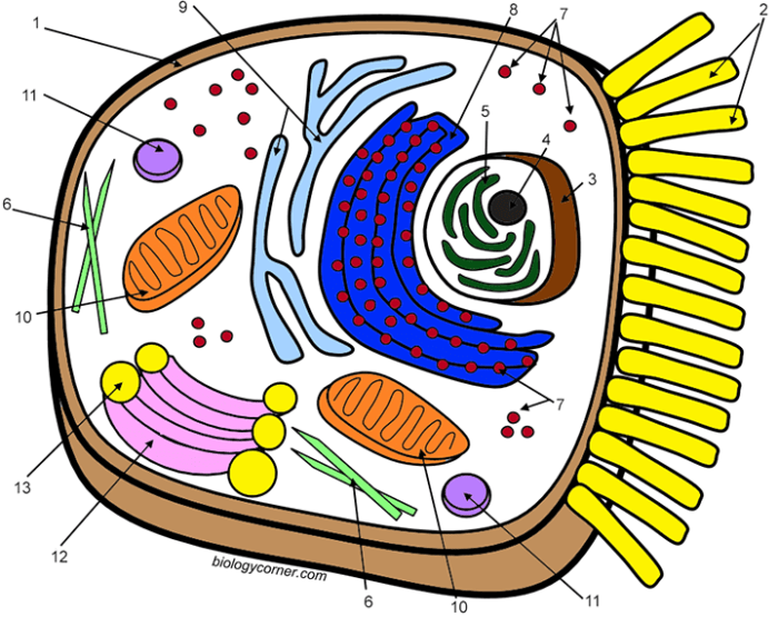
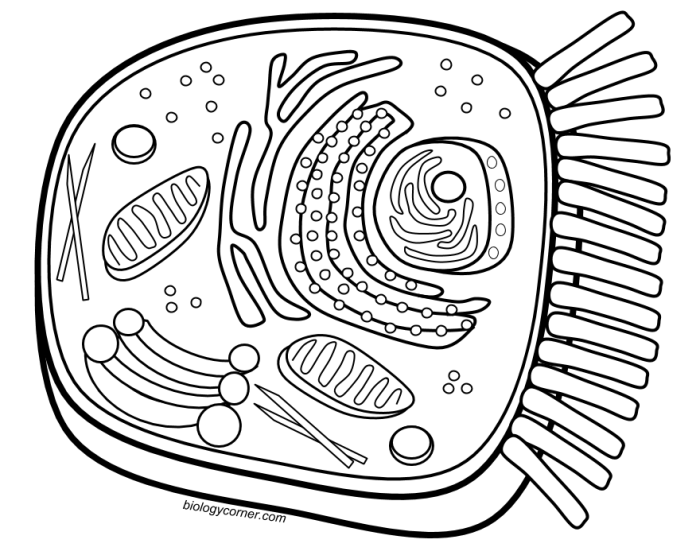
Animal cell coloring colored – The animal cell, a fundamental unit of life, is a marvel of intricate organization. Its internal architecture, a complex interplay of organelles, dictates its function and survival. Understanding these components is key to appreciating the dynamism and complexity of life itself. Each organelle contributes to the overall health and performance of the cell, working in a coordinated and often interdependent manner.
The cell, a microcosm of life, is not simply a bag of chemicals; it is a highly organized structure with specialized compartments performing specific tasks. This compartmentalization allows for efficient metabolic processes and prevents conflicting reactions. The cell membrane, a crucial boundary, controls the flow of materials in and out, maintaining the cell’s integrity and internal environment. This delicate balance is essential for cellular function and survival.
Cell Membrane: Maintaining Cellular Integrity
The cell membrane, a selectively permeable barrier, is the defining boundary of the animal cell. Composed primarily of a phospholipid bilayer interspersed with proteins and cholesterol, it regulates the passage of substances into and out of the cell. This selective permeability is crucial; it allows essential nutrients to enter and waste products to exit, while preventing the entry of harmful substances.
The fluid mosaic model describes this dynamic structure, where components move laterally within the membrane, constantly shifting and adapting. This flexibility allows the cell to respond to its environment and maintain its integrity. The membrane’s role is not merely passive; it actively participates in cell signaling and communication with other cells.
Major Organelles and Their Functions
The animal cell houses a variety of organelles, each with a specific role in maintaining cellular function. The nucleus, often described as the cell’s control center, houses the genetic material (DNA). The endoplasmic reticulum (ER), a network of interconnected membranes, synthesizes proteins and lipids. The Golgi apparatus processes and packages proteins for transport. Mitochondria, often referred to as the “powerhouses” of the cell, generate energy through cellular respiration.
Lysosomes contain enzymes that break down waste materials. The cytoskeleton provides structural support and facilitates cell movement. Ribosomes are responsible for protein synthesis. The centrosome organizes microtubules and plays a role in cell division.
| Organelle | Location | Function |
|---|---|---|
| Nucleus | Center of the cell | Houses DNA, controls gene expression |
| Cell Membrane | Outer boundary of the cell | Regulates passage of substances, maintains cell integrity |
| Endoplasmic Reticulum (ER) | Network of membranes throughout the cytoplasm | Protein and lipid synthesis |
| Golgi Apparatus | Near the ER | Processes and packages proteins |
| Mitochondria | Throughout the cytoplasm | Cellular respiration, ATP production |
| Lysosomes | Throughout the cytoplasm | Waste breakdown |
| Ribosomes | Free in cytoplasm or bound to ER | Protein synthesis |
| Cytoskeleton | Throughout the cytoplasm | Structural support, cell movement |
| Centrosome | Near the nucleus | Organizes microtubules, role in cell division |
Animal vs. Plant Cells: Structural and Functional Differences
While both animal and plant cells are eukaryotic, possessing membrane-bound organelles, they exhibit key differences. Plant cells possess a rigid cell wall, providing structural support and protection, absent in animal cells. Chloroplasts, responsible for photosynthesis, are found in plant cells but not in animal cells. Plant cells typically have a large central vacuole for storage and turgor pressure maintenance, a feature less prominent in animal cells.
These differences reflect the distinct lifestyles and functions of these two cell types. The presence of a cell wall in plant cells contributes to their ability to withstand osmotic stress, a challenge animal cells face differently through other mechanisms.
Coloring Techniques for Animal Cells
The vibrant world of the microscopic, often hidden from our naked eye, reveals itself through the careful application of color. Staining techniques, far from being mere aesthetic exercises, are crucial tools for understanding the intricate architecture and function of the animal cell. They allow us to distinguish between different cellular components, revealing a hidden complexity that informs our understanding of life itself.
The process, however, requires precision and a delicate touch, a dance between science and artistry.Preparing an animal cell slide for microscopic observation involves a series of meticulous steps, each essential for obtaining a clear and informative image. A failure at any stage can compromise the entire process, resulting in blurry, uninterpretable results. The precision required is akin to preparing a delicate miniature painting, where each brushstroke must be carefully considered.
Preparing Animal Cell Slides for Microscopic Observation
The preparation begins with obtaining a sample of animal cells. This could be a cheek swab, a blood sample, or a tissue culture. The sample is then gently spread onto a clean microscope slide. A drop of isotonic saline solution is added to prevent cell shrinkage or lysis. The slide is then carefully air-dried to fix the cells, ensuring they adhere to the surface.
The intricate detail of an animal cell, colored with vibrant hues under the microscope, reminded me of another kind of artistry. The precision is similar to the meticulous work found in animal coloring book pages , each stroke bringing a creature to life. Returning to the cell, I see now the same potential for creative expression, a miniature world waiting to be explored through color.
Once dried, the slide is ready for staining. The quality of the slide preparation directly impacts the clarity and detail observable under the microscope. A poorly prepared slide can obscure cellular structures, rendering the staining process ineffective.
Staining Methods for Animal Cells
Three common staining methods offer distinct advantages and disadvantages: simple staining, differential staining, and vital staining. Each method provides unique insights into cellular structures, depending on the specific goals of the observation. The choice of method depends critically on the research question and the type of information sought.
- Simple Staining: This method involves using a single stain, typically a basic dye like methylene blue, to color the entire cell. The advantage is its simplicity and speed. The disadvantage is its limited ability to differentiate between different cellular components. All structures appear similarly stained, making it difficult to distinguish the nucleus from the cytoplasm, for example.
- Differential Staining: This technique employs two or more stains to differentiate between different cellular components. Gram staining, for example, distinguishes between Gram-positive and Gram-negative bacteria. In animal cells, this approach can highlight specific organelles or structures, offering a more detailed view of cellular organization.
- Vital Staining: This method uses dyes that selectively stain living cells without causing immediate harm. This allows for the observation of cellular processes in real-time, providing dynamic insights into cellular activity. However, the choice of vital stain is crucial, as some dyes can eventually affect cell viability.
Specific Stains and Their Effects
Methylene blue, a common simple stain, readily penetrates the cell membrane and binds to negatively charged components within the cell, such as nucleic acids. It stains the nucleus a deep blue, while the cytoplasm appears a lighter blue. Hematoxylin, on the other hand, is a more complex stain that binds to negatively charged molecules, particularly in the nucleus, staining it a dark purple or blue-black.
It also stains the cytoplasm, but less intensely. The contrast between the stained nucleus and the cytoplasm allows for clear visualization of nuclear structure.
Visual Representation of a Methylene Blue Stained Animal Cell
Imagine a cell, roughly circular in shape. The nucleus, centrally located, is a vibrant, deep blue, its chromatin faintly visible as slightly darker specks within the intense blue. The cytoplasm surrounding the nucleus is a lighter, softer blue, almost a hazy watercolor wash compared to the nucleus’s bold hue. Small, darker blue dots, representing the ribosomes, are scattered throughout the lighter blue cytoplasm.
The cell membrane itself is not distinctly visible with this simple stain, appearing as a subtle change in the intensity of the blue at the cell’s periphery. The overall effect is one of stark contrast, a miniature cosmos of blue hues revealing the basic architecture of the animal cell.
Interpreting Colored Animal Cell Images: Animal Cell Coloring Colored

The vibrant hues of a stained animal cell slide, far from mere aesthetic appeal, offer a window into the intricate workings of life itself. The careful interpretation of these colors, their intensities, and their distributions is crucial for understanding cellular structure and function. A seemingly chaotic swirl of color reveals a precise choreography of organelles, each playing its vital role within the cell’s microscopic drama.The visual information gleaned from stained preparations allows for a deeper understanding beyond simple identification.
It provides insights into the relative abundance of specific organelles, their metabolic activity, and potential abnormalities. This interpretive process, however, requires a nuanced understanding of staining techniques and the potential for artifacts to cloud the interpretation.
Key Organelles Visible in Stained Animal Cell Images
A typical stained animal cell, viewed under a light microscope, might reveal several key organelles. The nucleus, often stained a deep purple or blue with hematoxylin, stands out as a large, generally spherical structure, housing the cell’s genetic material. The cytoplasm, the jelly-like substance filling the cell, may appear a pale pink or light blue depending on the counterstain used.
Within this cytoplasmic matrix, the endoplasmic reticulum might be visible as a network of interconnected tubules and sacs, often staining differently depending on whether it is rough (studded with ribosomes) or smooth. Ribosomes themselves, though individually too small to resolve, contribute to the overall staining intensity of the rough endoplasmic reticulum. Mitochondria, the cell’s powerhouses, might appear as small, rod-shaped structures, often stained a different color from the surrounding cytoplasm, perhaps a reddish-purple or even a darker blue depending on the specific dye.
Lysosomes, smaller and more numerous, may be difficult to distinguish definitively without specialized staining techniques. The Golgi apparatus, involved in protein modification and transport, might appear as a stacked series of flattened sacs, although its visualization often depends on the specific staining method. Finally, depending on the cell type and the staining protocol, vacuoles, often appearing as clear or lightly stained vesicles, may be visible.
Comparison of Organelle Appearance Under Different Staining Techniques
The appearance of organelles varies significantly depending on the staining technique employed. Hematoxylin and eosin (H&E) staining, a common histological stain, typically renders the nucleus a deep purple (hematoxylin) and the cytoplasm a pale pink (eosin). However, other stains, such as Giemsa or Wright stains, commonly used in hematology, produce different color palettes and highlight different cellular components. For instance, mitochondria might appear more prominently stained with certain dyes designed to target specific mitochondrial enzymes or membrane components, allowing for better visualization and potential assessment of mitochondrial activity.
The choice of stain directly impacts the contrast and visibility of different organelles, making the selection crucial for the specific research question. For example, a stain specific for lipid droplets would highlight these structures in adipocytes, which would not be readily apparent with H&E staining.
Color Intensity as an Indicator of Concentration or Activity
The intensity of staining reflects, to a degree, the concentration or activity level of the stained cellular component. A darkly stained nucleus might indicate a high concentration of DNA, perhaps reflecting a cell actively engaged in transcription or replication. Similarly, intensely stained mitochondria might suggest a high metabolic rate, while a pale staining might indicate reduced activity or damage.
However, it’s crucial to remember that staining intensity is not a direct, quantitative measure. Factors such as dye penetration, fixation artifacts, and the inherent variability of staining procedures all influence the final result. Therefore, careful controls and standardized protocols are essential for reliable interpretation. For instance, comparing the mitochondrial staining intensity across a population of cells under different experimental conditions can provide insights into changes in metabolic activity.
Potential Artifacts in Stained Animal Cell Preparations, Animal cell coloring colored
Several artifacts can arise during the preparation of stained animal cell slides, potentially leading to misinterpretations. These artifacts include precipitation of the stain itself, creating spurious dark spots; shrinkage or distortion of the cells due to fixation or dehydration; the presence of air bubbles creating clear spaces within the preparation; and the formation of precipitates from the fixative or stain.
Furthermore, uneven staining can result from inadequate penetration of the stain or variations in the cell’s permeability. Careful attention to detail during sample preparation and staining, along with awareness of these potential artifacts, is essential for accurate interpretation of the results. For example, a careful examination of the background staining can help distinguish between true cellular structures and staining precipitates.
Applications of Animal Cell Coloring
The vibrant hues revealed through cell staining are not merely aesthetic; they are crucial tools unlocking profound insights into the intricate world of animal cells. From the clinical setting to the research laboratory, the application of color to these microscopic building blocks provides a window into health, disease, and the very mechanisms of life. The ability to visualize specific cellular structures and processes is transformative, impacting diagnosis, treatment, and our fundamental understanding of biology.The precise application of color to cells, a technique refined over centuries, allows for the identification of otherwise invisible structures and processes.
This visual clarity is paramount in various fields, enabling researchers and medical professionals to diagnose diseases, track cellular responses to treatments, and advance scientific knowledge.
Cell Staining in Medical Diagnosis
Microscopic examination of stained cells forms the bedrock of many medical diagnoses. For instance, Pap smears, utilizing stains like Papanicolaou stain, allow for the early detection of cervical cancer by highlighting abnormal cellular changes. Similarly, blood smears stained with Wright-Giemsa stain enable the identification of various blood cell types and abnormalities, aiding in the diagnosis of anemias, leukemia, and other hematological disorders.
The distinct coloration of different cellular components—nuclei, cytoplasm, and organelles—provides crucial diagnostic information, often leading to timely interventions and improved patient outcomes. The specificity of the staining process allows for the clear identification of cancerous cells, infectious agents, and other pathological indicators, significantly enhancing diagnostic accuracy.
Colored Animal Cell Images in Research and Education
Colored images of animal cells are indispensable tools in both research and educational settings. In research, these images serve as primary data, providing visual evidence of experimental results. For example, immunofluorescence microscopy, utilizing fluorescently labeled antibodies, creates vividly colored images showcasing the localization of specific proteins within cells. This technique is vital for understanding cellular pathways, protein interactions, and the effects of various treatments.
In education, colorful images significantly enhance learning and understanding. Textbooks, presentations, and online resources rely heavily on such images to illustrate complex cellular structures and processes, making abstract concepts more accessible and engaging for students. The visual clarity provided by these images is paramount in conveying the intricacies of cell biology.
Revealing Specific Cellular Processes Through Staining Techniques
Different staining techniques highlight specific aspects of cellular function. For example, hematoxylin and eosin (H&E) staining, a common histological technique, stains nuclei blue and cytoplasm pink, providing a general overview of tissue architecture and cell morphology. However, more specialized techniques reveal much finer details. Immunohistochemistry (IHC) utilizes antibodies conjugated to enzymes or fluorescent molecules to visualize specific proteins, providing insights into protein expression and localization.
This technique is crucial in cancer research, allowing for the identification of tumor markers and the assessment of treatment efficacy. Similarly, techniques like Golgi staining selectively highlight the Golgi apparatus, revealing its structure and function within the cell. The choice of staining technique directly dictates the information obtained, demonstrating the power of targeted visualization in cell biology.
Scientific Applications of Animal Cell Coloring
The importance of animal cell coloring extends far beyond basic observation. The following list showcases diverse scientific applications:
- Cancer Diagnosis and Treatment Monitoring: Staining techniques are used to identify cancerous cells, assess tumor grade, and monitor the effectiveness of cancer therapies.
- Infectious Disease Diagnosis: Staining is crucial for identifying pathogens like bacteria and parasites in infected tissues.
- Neurobiology Research: Specific stains help visualize neuronal structures and connections, aiding in the study of the nervous system.
- Drug Development and Testing: Cell staining allows researchers to assess the effects of new drugs on cellular processes.
- Genetic Research: Techniques like fluorescence in situ hybridization (FISH) utilize fluorescent probes to visualize specific DNA sequences within cells, aiding in genetic analysis and diagnosis.
Q&A
What safety precautions should I take when handling stains?
Always wear appropriate personal protective equipment (PPE), including gloves and eye protection, when handling stains. Work in a well-ventilated area and follow the manufacturer’s safety instructions.
How long does it typically take to stain an animal cell sample?
Staining time varies depending on the stain and protocol used, ranging from minutes to hours. Consult your chosen protocol for specific timing instructions.
What are some common mistakes to avoid when staining animal cells?
Common mistakes include over-staining, insufficient staining, improper slide preparation, and using expired reagents. Careful adherence to protocols minimizes errors.
Can I use household dyes to stain animal cells?
No, household dyes are not suitable for staining animal cells for scientific purposes. They lack the specificity and reliability of biological stains.

