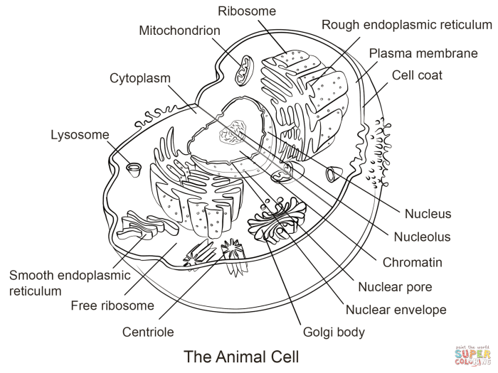
The Role of Pigments in Animal Cell Color: Colored Animal Cell Coloring

Colored animal cell coloring – So, you’ve colored your animal cells, but have you ever wonderedwhy* they’re so gosh-darned colorful? It’s not just for artistic flair, my friend! The vibrant hues are all thanks to the amazing world of pigments, the tiny molecular artists within our cells. Let’s dive into the fascinating chemistry of color!
Pigments are essentially molecules that absorb certain wavelengths of light and reflect others. The wavelengths that are reflected are the ones we perceive as color. Think of it like this: a red pigment absorbs all the colors except red, which it bounces back at your eyeballs. Pretty neat, huh? This absorption and reflection is all down to the pigment’s chemical structure – the arrangement of atoms and the types of bonds between them influence how they interact with light.
Types of Animal Cell Pigments and Their Chemical Structures
Pigments in animal cells come in a variety of flavors, each with its own unique chemical makeup and resulting color. Melanin, for example, is a family of pigments responsible for the brown, black, and reddish hues in many animals. Its structure is a complex polymer, meaning it’s a long chain of smaller molecules linked together. The exact structure varies, resulting in the range of colors we see.
Another major player is carotenoid, which gives many animals their yellow, orange, and red tones. These pigments are built from isoprene units, arranged in specific chains that determine their color. Lastly, we have haemoglobin, the star of the red blood cell show, which contains iron and is responsible for the crimson color. Its complex structure involves a heme group with an iron atom at its center, allowing it to bind oxygen and give blood its characteristic color.
Examples of Animal Pigments and Their Corresponding Animals
Let’s look at some real-world examples to make this less abstract and more, well, colorful!
The following list highlights the diverse range of animal pigments and their effects on animal coloration.
- Melanin: Found in humans, giving us our skin, hair, and eye color. Think of all the beautiful shades of brown, black, and even reddish hues! A chameleon’s color-changing abilities are also partially due to melanin.
- Carotenoids: These give flamingos their pink plumage and carrots their orange color. Many fish and birds also owe their vibrant hues to these pigments. Think of the brilliant orange of a goldfish or the bright yellow of a canary.
- Haemoglobin: The star of the show in red blood cells, responsible for that vital crimson color. It’s what makes our blood, and the blood of many other animals, red.
Light Absorption Properties of Different Pigments
Different pigments have different affinities for various wavelengths of light.
This means some absorb more strongly in certain areas of the visible spectrum than others. For instance, melanin absorbs broadly across the visible spectrum, leading to its dark colors. Carotenoids, on the other hand, typically absorb blue and green light more strongly, reflecting yellow, orange, and red light. Haemoglobin, with its unique iron-containing structure, absorbs most wavelengths except red, which is strongly reflected.
This explains the color differences seen in various animals. The interplay of these different pigments and their absorption properties creates the stunning array of colors we see in the animal kingdom.
Understanding colored animal cell coloring provides a foundational visual understanding of cellular structures. To further explore animal biology visually, consider using a resource like the animal anatomy coloring book which offers a broader perspective on animal systems. Returning to the cellular level, colored animal cell coloring helps solidify knowledge of organelles and their functions within the complex animal body.
Techniques for Visualizing Colored Animal Cells

So, you’ve got yourself some gloriously colored animal cells, and now you want to show them off? Excellent! Let’s dive into the exciting world of microscopy and cell staining – because let’s face it, nobody wants to see a boring, colorless cell. We’re aiming for vibrant, show-stopping cell visuals here!
Visualizing the colorful world of animal cells requires a bit more than just a magnifying glass (although, that’s a good start!). Different microscopy techniques, combined with clever staining methods, are our secret weapons in this quest for cellular beauty. Think of it as a cellular makeover, but with science!
Microscopy Techniques for Observing Animal Cell Coloration
Several microscopy techniques can reveal the stunning hues of animal cells. The choice depends on the level of detail and the specific colors you’re interested in. Let’s explore some popular options, shall we?
Light Microscopy: The workhorse of cell visualization. It’s relatively simple and affordable, making it a great starting point. However, light microscopy has its limitations; it can’t resolve structures smaller than the wavelength of visible light. Still, for observing the overall color and morphology of many colored cells, it’s a fantastic tool. Imagine seeing the vibrant orange of a carotenoid-rich cell under a light microscope – truly mesmerizing!
Fluorescence Microscopy: This technique uses fluorescent dyes or proteins that emit light at a specific wavelength when excited by a light source. This allows for incredibly specific visualization of particular cellular components, even if they’re not inherently colored. For example, you could use fluorescent antibodies to target and highlight specific proteins within a cell, making their location and distribution easily visible against a background of different colors.
Confocal Microscopy: A more advanced version of fluorescence microscopy, confocal microscopy uses lasers to scan the sample and create incredibly sharp, high-resolution images. It minimizes background noise and allows for 3D imaging of cells, revealing intricate details of color distribution within the cell. Picture a stunning 3D reconstruction of a cell, with each colored organelle perfectly defined – a true work of art!
Preparation Procedures for Visualizing Colored Cells Using Light Microscopy
Getting your cells ready for their close-up requires careful preparation. Think of it as prepping a celebrity for a photoshoot – the better the preparation, the better the results!
- Sample Preparation: Gently collect your colored cells (e.g., using a pipette or scraping). Avoid crushing them – we want to see those beautiful colors intact!
- Mounting: Place a small drop of your cell suspension onto a clean microscope slide. Add a coverslip carefully to avoid air bubbles. Air bubbles are the enemy of beautiful microscopy.
- Microscope Adjustment: Adjust the focus and light intensity on your microscope to achieve optimal viewing conditions. Experiment with different lighting settings to highlight the colors of your cells.
Step-by-Step Protocol for Staining Animal Cells, Colored animal cell coloring
Sometimes, even naturally colored cells need a little help to really pop. That’s where staining comes in. This is where the fun (and the slightly messy) part begins!
- Fixation: Gently fix your cells using a suitable fixative (e.g., formaldehyde) to preserve their structure and color.
- Staining: Apply the chosen stain (e.g., hematoxylin and eosin, or specialized stains for specific cellular components). Follow the manufacturer’s instructions carefully – we don’t want any accidental explosions of color!
- Washing: Rinse the cells thoroughly with water or buffer to remove excess stain.
- Mounting: Mount the stained cells onto a microscope slide and add a coverslip, as described earlier. Avoid bubbles!
- Microscopy: Observe your vibrantly stained cells under the microscope! Marvel at the intensified colors and the enhanced detail.
Examples of Animal Cell Types and Their Color Characteristics
Let’s look at some examples of naturally colored cells and how their unique pigments contribute to their appearance. These descriptions are purely imaginative, as I cannot provide actual images.
Example 1: A vibrant orange pigment cell from a tropical fish: Imagine a cell bursting with a rich, deep orange hue. This intense color is due to carotenoid pigments, concentrated within the cell’s cytoplasm. Under a microscope, you’d see tiny droplets of orange pigment dispersed throughout the cell, giving it its vibrant appearance. The cell’s shape is elongated, with a slightly flattened appearance.
Example 2: A reddish-brown pigment cell from a chameleon: Picture a cell exhibiting a complex pattern of reddish-brown hues. These colors arise from melanins, pigments that are distributed unevenly within the cell, creating a mottled effect. The cell itself is irregular in shape, with numerous small projections extending from its surface.
Example 3: A bright blue pigment cell from a blue morpho butterfly: Envision a cell with an almost iridescent blue color. This striking hue isn’t from a pigment but rather from the structural organization of tiny scales on the butterfly’s wings. These scales reflect light in a specific way, creating the brilliant blue effect. Under a microscope, you’d see these scales arranged in a highly ordered pattern.
FAQ Guide
What are some common challenges in visualizing colored animal cells?
Challenges include achieving optimal staining without damaging cells, distinguishing between natural and artificial colors, and resolving the fine details of cellular structures under the microscope.
How does cell coloration relate to animal health?
Abnormal cell coloration can indicate disease or cellular dysfunction. Changes in pigment production can signal problems with metabolic pathways or genetic abnormalities.
Can environmental factors permanently alter cell coloration?
While some changes are temporary, others, particularly those affecting genetic expression, can lead to permanent alterations in cell coloration, passed down through generations.
