Educational Applications of the Coloring Page

This animal cell coloring page offers a fun and engaging way to introduce complex biological concepts to young learners. Its versatility allows for diverse applications across various educational settings, significantly enhancing understanding and retention. The visual nature of the activity caters to different learning styles, making it an effective tool for both classroom and homeschool environments.Classroom Activities Using the Coloring PageThis coloring page can be integrated into various classroom activities.
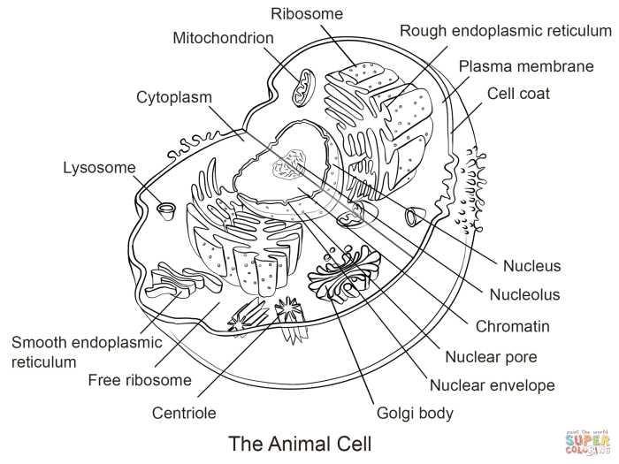
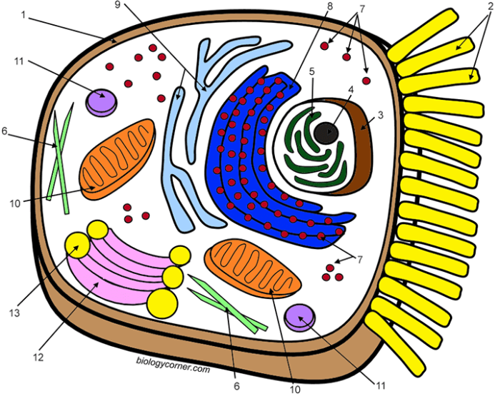
For example, it can be used as a pre-lesson activity to gauge prior knowledge and spark curiosity about cell structures. Following a lecture or presentation on animal cells, the coloring page can serve as a reinforcement activity, allowing students to actively apply what they’ve learned. Finally, it can be incorporated into a larger project, such as creating a classroom-wide cell model display, where students collaborate and share their knowledge.Enhancing Understanding of Cell Structure Through ColoringThe act of coloring the different organelles of the animal cell – the nucleus, mitochondria, ribosomes, and more – helps children visualize their relative sizes, locations, and functions.
By labeling each organelle as they color, students actively engage with the terminology and concepts, improving their understanding and memorization. The visual representation created through coloring aids in the formation of a mental model of the cell, a crucial step in comprehending its complexity. This hands-on approach allows for a deeper understanding than simply reading about cell structures from a textbook.Benefits of Hands-On Learning in Science EducationHands-on learning activities, such as coloring pages, are demonstrably beneficial for science education.
They transform abstract concepts into tangible experiences, making learning more engaging and memorable. The tactile nature of coloring fosters active participation, increasing focus and concentration. Furthermore, the collaborative potential of such activities promotes teamwork and communication skills, crucial aspects of scientific inquiry. Studies have shown that students who participate in hands-on activities often demonstrate improved understanding and retention of scientific concepts compared to those who rely solely on passive learning methods.
For instance, a study published in the Journal of Educational Psychology showed a significant improvement in students’ understanding of the human circulatory system after participating in a hands-on model-building activity.
Educational materials like animal cell coloring pages offer a hands-on approach to learning biology. For those seeking a creative outlet after completing their science studies, a wide variety of options exist, including anime coloring pages free for a fun and relaxing activity. Returning to the scientific realm, accurately colored animal cell diagrams can solidify understanding of cellular structures and processes.
Illustrative Examples of Animal Cell Components

Yo, Makassar crew! Let’s dive deep into the awesome world of animal cells. This coloring page isn’t just about crayons; it’s about understanding the tiny powerhouses that make up all of us (and every animal!). We’re gonna break down some key components, so grab your pencils and let’s get creative.
The Nucleus: The Cell’s Boss, Animal cell coloring page
Think of the nucleus as the brain of the cell – the control center. In your coloring page, depict it as a large, roundish structure, usually located near the center. It should be a darker color than the rest of the cell to emphasize its importance. You can add a slightly speckled texture inside to represent the chromatin (DNA) that holds all the cell’s genetic information.
Imagine it as a densely packed library holding all the blueprints for building and running the cell.
Mitochondria: The Power Plants
These are the energy factories! Show them as bean-shaped or sausage-shaped structures scattered throughout the cell. Give them a slightly textured look, maybe with some inner folds or cristae (that’s what they’re called!). Use a different color from the nucleus, something vibrant like red or purple, to show they’re the energetic heart of the cell. They’re like tiny power plants constantly generating energy for all the cell’s activities.
Ribosomes: The Tiny Protein Factories
Ribosomes are super small, so represent them as tiny dots or small ovals scattered throughout the cytoplasm (the jelly-like stuff filling the cell). You can cluster some around the endoplasmic reticulum (we’ll get to that next!), since that’s where many of them work. Use a different color again, maybe a light blue or green, to make them stand out from the other cell components.
These are the protein-making machines – essential for all cell functions.
The Cell Membrane: The Protective Barrier
Visualize the cell membrane as a thin, continuous line outlining the entire cell. It’s like a flexible, protective wall that controls what enters and exits the cell. You can draw it as a slightly wavy line, not perfectly smooth, to show its flexibility. It’s selectively permeable, meaning it carefully chooses what can pass through – like a super-strict bouncer at a club.
Think of it as a two-layered structure, representing the lipid bilayer – a double layer of fats that forms the basis of the membrane.
Endoplasmic Reticulum and Golgi Apparatus: The Cell’s Shipping and Receiving
The endoplasmic reticulum (ER) is a network of interconnected tubes and sacs. For simplicity, represent it as a network of interconnected lines within the cell, spreading out from the nucleus. You can color it a different shade from the cytoplasm. The rough ER (with ribosomes attached) could have small dots on it representing the ribosomes. The Golgi apparatus can be shown as a stack of flattened sacs or pancakes near the ER.
It receives, modifies, and packages proteins made by the ribosomes for delivery throughout the cell or outside of it – the cell’s shipping and receiving department. Color it distinctly from the ER, maybe a different shade of the same color, to show its distinct function.
Variations and Extensions of the Coloring Page: Animal Cell Coloring Page
This section explores different ways to adapt the animal cell coloring page to suit various age groups and learning styles, enhancing engagement and understanding. We’ll present three variations with increasing complexity, alongside suggestions for interactive elements to make the learning experience more dynamic and fun, bro, like adding a bit of Makassar spice to your biology lesson!
By offering varying levels of complexity, we cater to different learning paces and abilities, ensuring everyone gets a chance to “boss” the cell structure. Interactive elements further solidify understanding and turn a simple coloring activity into an engaging learning adventure.
Animal Cell Coloring Page Variations
The following table Artikels three variations of the animal cell coloring page, categorized by complexity and suggested age range. Each variation offers a progressively challenging experience, building upon the foundational knowledge gained in simpler versions. Think of it as leveling up your cell knowledge, from newbie to pro!
| Complexity Level | Age Range | Description | Interactive Elements |
|---|---|---|---|
| Beginner | 5-7 years | A simplified diagram showing only the major organelles (nucleus, cell membrane, cytoplasm). Organelles are large and easily colored. Minimal detail is included, focusing on basic shapes and colors. Think of it as a basic introduction, like learning the alphabet before writing a novel. | Color-by-number activity where each organelle is assigned a number and a corresponding color. |
| Intermediate | 8-10 years | Includes more organelles (mitochondria, ribosomes, Golgi apparatus, endoplasmic reticulum). Organelles are more realistically proportioned and detailed. It’s like adding more instruments to your band – more complex, but more rewarding. | Labeling activity with a word bank provided. Students match the organelle names to their locations on the diagram. |
| Advanced | 11+ years | A detailed diagram showing all major and minor organelles, with accurate representations of their structures and relative sizes. This version challenges students to understand the intricate relationships between organelles. It’s the full orchestra playing a symphony! | Fill-in-the-blank activity where students describe the function of each organelle in their own words. This pushes them to really understand what’s going on inside the cell. |
Top FAQs
What type of paper is best for printing an animal cell coloring page?
Heavier weight paper, like cardstock, is ideal for durability and to prevent bleed-through from markers or colored pencils.
Can I use digital coloring tools instead of traditional crayons?
Absolutely! Many digital art programs and apps allow you to color the page digitally for a modern twist.
How can I make the coloring page more interactive?
Include blank labels for students to name the organelles, or add a simple quiz on the back related to the cell’s functions.
Where can I find free animal cell images to use as inspiration?
A quick search on royalty-free image sites or educational websites will provide plenty of inspiration for your design.

