Worksheet Overview

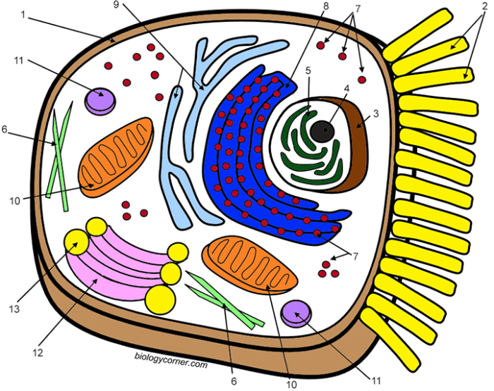
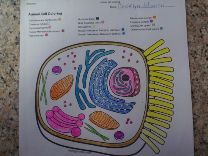
Animal cell coloring worksheet answer key – Animal cell coloring worksheets are educational tools designed to help students visualize and learn the structures and functions of various organelles within an animal cell. These worksheets typically present a simplified diagram of an animal cell, often with numbered or labeled sections corresponding to specific organelles. Students then color-code these sections according to a key or instructions, reinforcing their understanding of cell structure.
The visual nature of the activity makes it particularly effective for younger learners or those who benefit from visual aids.Animal cell coloring worksheets commonly include a range of organelles, each playing a crucial role in the cell’s overall function. Understanding these organelles and their roles is fundamental to comprehending cellular biology. The level of detail included varies depending on the age and knowledge level of the intended audience.
Organelles Commonly Included in Animal Cell Coloring Worksheets and Their Functions
The following table details several organelles frequently featured in animal cell coloring worksheets, along with their functions and typical representations in such worksheets. The colors suggested are common choices but may vary across different worksheets.
| Organelle | Function | Shape | Example Color |
|---|---|---|---|
| Nucleus | Contains the cell’s genetic material (DNA) and controls cell activities. | Large, spherical | Purple or dark blue |
| Cell Membrane | Encloses the cell, regulating the passage of substances in and out. | Thin, flexible boundary | Light blue or black Artikel |
| Cytoplasm | The jelly-like substance filling the cell, containing organelles. | Fills the cell | Light yellow or beige |
| Mitochondria | Produce energy (ATP) through cellular respiration. | Oval or rod-shaped | Red or pink |
| Ribosomes | Synthesize proteins. | Small dots | Dark brown or grey |
| Endoplasmic Reticulum (ER) | Network of membranes involved in protein and lipid synthesis and transport. (Rough ER has ribosomes attached; Smooth ER does not). | Network of interconnected tubes and sacs | Light green (Rough ER), Light orange (Smooth ER) |
| Golgi Apparatus (Golgi Body) | Processes and packages proteins and lipids for transport. | Stack of flattened sacs | Yellow or light orange |
| Lysosomes | Break down waste materials and cellular debris. | Small, spherical sacs | Dark green or purple |
| Vacuoles | Store water, nutrients, and waste products. Generally smaller and more numerous in animal cells than plant cells. | Small, membrane-bound sacs | Light blue or clear |
Organelle Identification and Function

Identifying animal cell organelles from visual representations presents unique challenges for students. Microscopic images often lack sharp contrast, making it difficult to distinguish the boundaries and fine details of various organelles. Furthermore, the size and appearance of organelles can vary depending on the cell type, its stage in the cell cycle, and the preparation techniques used for microscopy.
This can lead to confusion and misidentification, hindering a thorough understanding of cellular structure and function.
Challenges in Organelle Identification
Visual identification of organelles relies heavily on students’ ability to interpret microscopic images. The small size and overlapping nature of organelles in a crowded cellular environment create significant obstacles. For instance, distinguishing between the endoplasmic reticulum (ER) and the Golgi apparatus can be difficult due to their similar appearance in some images. Similarly, lysosomes and peroxisomes, both small membrane-bound organelles, may be challenging to differentiate based solely on their visual characteristics.
The lack of distinct visual markers for certain organelles further complicates identification. Moreover, variations in staining techniques used in microscopy can affect the visibility and appearance of organelles, adding another layer of complexity.
Comparative Table: Animal and Plant Cells
A comparative analysis of animal and plant cells highlights the unique characteristics of each. The following table illustrates the presence or absence of key organelles:
| Organelle | Animal Cell | Plant Cell | Description of Difference |
|---|---|---|---|
| Cell Wall | Absent | Present | Provides structural support and protection in plant cells. |
| Chloroplasts | Absent | Present | Sites of photosynthesis, converting light energy into chemical energy. |
| Large Central Vacuole | Absent or small | Present | Maintains turgor pressure, stores water and nutrients in plant cells. |
| Plasmodesmata | Absent | Present | Channels that connect adjacent plant cells, facilitating communication and transport. |
| Centrioles | Present | Usually absent | Play a role in cell division, organizing microtubules. Some lower plant species may have them. |
Strategies for Effective Organelle Differentiation
Effective teaching strategies are crucial for helping students overcome the challenges of identifying and differentiating cell organelles. Combining multiple learning modalities can significantly improve understanding. For example, integrating interactive 3D models alongside traditional microscopic images allows for a more comprehensive visualization of organelle structure and spatial relationships within the cell. Furthermore, incorporating labeled diagrams and flowcharts that summarize the key functions of each organelle can aid in memorization and comprehension.
Guided practice activities, such as matching games or quizzes that require students to identify organelles based on their characteristics, can reinforce learning and assess understanding. Finally, encouraging students to actively compare and contrast the functions of different organelles helps to solidify their knowledge and improve their ability to differentiate between them.
Answer Key Development and Considerations: Animal Cell Coloring Worksheet Answer Key
Creating a comprehensive and user-friendly answer key is crucial for the success of any educational worksheet. A well-designed key not only provides correct answers but also aids in understanding the underlying concepts and prevents the propagation of misconceptions. This section details the development of an answer key for a hypothetical animal cell coloring worksheet, emphasizing clarity, accuracy, and ease of use for both educators and students.The accuracy of the answer key is paramount.
Inaccurate information can lead to significant learning deficits and reinforce incorrect understandings of cell biology. Therefore, careful attention must be paid to the correct identification and description of each organelle and its function. The chosen colors should also be visually distinct to facilitate easy identification and memorization. The answer key’s design should minimize ambiguity, enabling both teachers and students to quickly and accurately verify the completed worksheet.
Hey there, champ! Finished your animal cell coloring worksheet answer key? Feeling all scientific and clever? Well, why not switch gears a bit and check out this super fun farm animal coloring sheet for a delightful change of pace! It’s a great way to relax after all that cell studying. Then, get back to those amazing animal cells – you’re gonna ace it!
Animal Cell Coloring Worksheet Answer Key
The following table provides a hypothetical answer key for an animal cell coloring worksheet. Color suggestions are provided, but these can be adapted based on available materials and personal preferences. It is important to ensure color choices offer sufficient contrast for clarity.
| Organelle | Color Suggestion | Description |
|---|---|---|
| Cell Membrane | Dark Blue | The outer boundary of the cell, regulating the passage of substances. |
| Cytoplasm | Light Yellow | The jelly-like substance filling the cell, containing organelles. |
| Nucleus | Purple | The control center of the cell, containing genetic material (DNA). |
| Nucleolus | Darker Purple | Located within the nucleus; involved in ribosome production. |
| Ribosomes | Small Red Dots | Sites of protein synthesis, found free in the cytoplasm or attached to the endoplasmic reticulum. |
| Endoplasmic Reticulum (Rough) | Light Green | Network of membranes studded with ribosomes; involved in protein synthesis and transport. |
| Endoplasmic Reticulum (Smooth) | Light Green (slightly different shade from rough ER) | Network of membranes lacking ribosomes; involved in lipid synthesis and detoxification. |
| Golgi Apparatus | Orange | Modifies, sorts, and packages proteins and lipids for secretion or transport. |
| Mitochondria | Dark Red | Powerhouses of the cell; generate ATP through cellular respiration. |
| Lysosomes | Dark Green | Contain digestive enzymes; break down waste materials and cellular debris. |
| Centrioles | Brown | Involved in cell division and organization of microtubules. |
Strategies for Creating a Clear and Understandable Answer Key
To ensure clarity and ease of use, the answer key should be well-organized and visually appealing. A tabular format, as shown above, is highly recommended. Each organelle should be clearly labeled, with a concise description of its function. Using color-coding consistent with the worksheet itself helps students quickly cross-reference their work. Furthermore, a key should be designed to avoid ambiguity; for instance, using distinct color shades for organelles with similar appearances (e.g., rough and smooth ER) prevents confusion.
Including visual aids, such as simplified diagrams showing the relative location of organelles, can further enhance understanding. The overall goal is to create a resource that is both informative and accessible to all users.
Visual Representation and Accuracy
Accurate visual representation of organelles in a coloring worksheet is crucial for effective learning. A well-executed worksheet reinforces understanding of organelle structure, relative size, and location within the cell, facilitating memorization and comprehension of cellular processes. Inaccurate depictions, conversely, can lead to misconceptions and hinder learning.The accuracy of a cell diagram directly impacts the student’s ability to visualize and internalize the complex relationships between different organelles.
A visually appealing and correctly structured worksheet can transform a potentially tedious task into an engaging and educational experience. Conversely, a worksheet with inaccurate or poorly drawn organelles can create confusion and frustration, undermining the educational objective.
Common Errors in Depicting Cell Organelles and Their Avoidance
Common errors in depicting animal cell organelles often involve inaccuracies in shape, size, and location. For instance, the nucleus is frequently drawn too small relative to the cell’s overall size, or the Golgi apparatus might be represented as a simple, uniform structure instead of its characteristic stacked cisternae. Mitochondria are sometimes depicted as simple ovals rather than their more complex, bean-shaped forms with inner cristae.
The endoplasmic reticulum (ER) is often simplified or omitted entirely, despite its extensive network within the cell. Lysosomes are frequently overlooked or drawn disproportionately small.To avoid these errors, detailed reference images from reputable sources (textbooks, scientific journals, or educational websites) should be consulted. Careful attention to the relative size and spatial arrangement of organelles is paramount. Using accurate templates or pre-drawn Artikels can aid in maintaining proportions and positioning.
Employing different colors for different organelles enhances visual distinction and aids in memorization. Finally, incorporating clear labeling can prevent confusion and further solidify learning.
A Correctly Colored Animal Cell
Imagine a circular cell, approximately 10-20 micrometers in diameter, with a large, centrally located nucleus occupying about 10% of the cell’s volume. The nucleus is colored a light purple, containing darker purple nucleoli. Surrounding the nucleus is a network of light pink endoplasmic reticulum (ER), with rough ER (studded with ribosomes, depicted as small dark blue dots) appearing more granular and concentrated near the nucleus, and smooth ER extending further into the cytoplasm.
Scattered throughout the cytoplasm are numerous small, dark blue ribosomes. The Golgi apparatus is represented as a series of stacked, flattened sacs (cisternae) colored light green, located near the nucleus but distinct from the ER. Numerous bean-shaped mitochondria, colored a vibrant red, are distributed throughout the cytoplasm, reflecting their role in energy production. Small, dark purple lysosomes are scattered sparsely across the cytoplasm.
The cell membrane, a thin, dark brown line, encloses the entire cell. The cytoplasm itself is a light yellow, providing a contrasting background for the various organelles. The relative sizes of the organelles are accurately represented, reflecting their actual proportions within a typical animal cell.
Frequently Asked Questions
What are some common mistakes students make when coloring animal cell organelles?
Common mistakes include inaccurate sizing of organelles, incorrect placement within the cell, and using inappropriate colors that don’t reflect the organelle’s function or typical representation.
How can I adapt this worksheet for different age groups?
Adapt by adjusting the level of detail and complexity. Younger students might focus on fewer organelles, while older students can explore more intricate structures and functions.
What alternative assessment methods can be used alongside the coloring worksheet?
Consider quizzes, short answer questions, labeling diagrams, creating 3D models, or presentations to assess understanding beyond the coloring activity.
Are there any online resources that can supplement the worksheet?
Many online resources offer interactive models, animations, and further information on animal cell structures and functions. A simple web search will yield many useful results.

