Coloring Techniques and Strategies for Accuracy

Labeled animal cell coloring – Yo, peeps! So you’re tackling an animal cell diagram, huh? Making it look legit isn’t just about slapping on some colors; it’s about showing you actuallyget* the cell’s vibe. We’re talking about accuracy, making those organelles pop, and showing how they all work together like a super-tight crew. Let’s get this coloring game on point.
Accurate representation of organelles is key to understanding their function. Using the right techniques and colors will make your diagram clear and easy to understand, helping you ace that biology test or just impress your friends with your mad cell-coloring skills. Think of it like this: a messy, inaccurate coloring job is like a blurry Instagram pic—no one’s gonna be impressed.
A clean, precise one? That’s the fire selfie that gets all the likes.
Color Choices for Organelle Differentiation
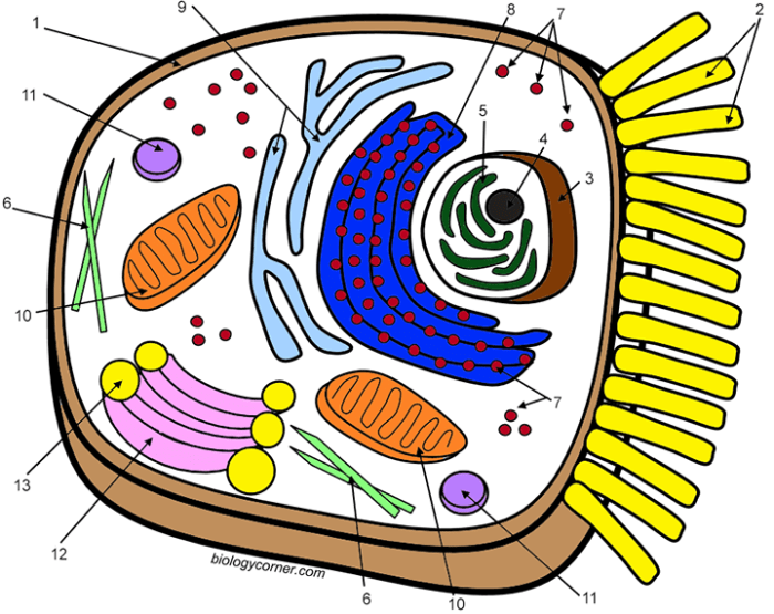
Choosing the right colors is crucial for making your organelles easily distinguishable. Think about contrast – you don’t want two organelles blending together like a bad mixtape. For example, the nucleus, being the boss of the cell, could be a bold dark purple. The mitochondria, the cell’s powerhouses, could be a vibrant red or orange, to show their energy-producing action.
The endoplasmic reticulum, with its network of tubes, could be a light blue or green to show its intricate structure. Using contrasting colors makes it a breeze to identify each organelle. Avoid pastel shades for main organelles; go for bold and distinct hues. Think less “watercolor painting” and more “street art” – make it pop!
Labeled animal cell coloring is a great way to learn biology, helping visualize the intricate structures within a cell. For a different creative outlet, you might enjoy the expressive style of female anime coloring pages , which offer a fascinating contrast to the scientific precision of cell diagrams. Returning to the scientific realm, remember to carefully label each organelle in your animal cell drawing for a complete understanding.
Using Color to Highlight Organelle Relationships
Colors aren’t just for showing differences; they can also show how organelles work together. For instance, you could use shades of the same color to illustrate organelles involved in a specific pathway. Let’s say you want to highlight the relationship between the ribosomes (which make proteins) and the endoplasmic reticulum (which transports proteins). You could color the ribosomes a dark blue and the ER a lighter blue, creating a visual connection that shows their teamwork.
This method is all about visual storytelling – showing how different parts of the cell interact.
Step-by-Step Guide for Coloring a Labeled Animal Cell Diagram
Alright, let’s break it down step-by-step. This ain’t rocket science, but it’s about precision:
- Sketch it out: Lightly sketch the cell and its organelles first. This helps you plan your color scheme and placement. Don’t go crazy with detail yet; just get the basic shapes down.
- Base Colors: Start with your base colors for each organelle. Remember those contrasting colors we talked about? This is where they shine. Use light, even strokes to avoid smudging.
- Adding Detail: Once your base colors are down, add details to your organelles. You can use shading or different shades of the same color to give them depth and dimension. Think about using darker shades for areas that are more shadowed or deeper within the cell.
- Labeling: Use a clear, easy-to-read font for your labels. Make sure your labels are neatly placed and clearly linked to the correct organelle. A different color for the labels is a good idea.
- Final Touches: Check for any smudges or areas that need more color. Make sure your colors are consistent and your labels are accurate. A little clean-up goes a long way!
Illustrative Examples of Labeled Animal Cells

Yo, peeps! Let’s dive into some seriously rad illustrations of animal cells. We’re gonna check out three different levels of detail, from super basic to full-on cellular bling. Think of it as leveling up your cell-knowledge game!We’ll break down each illustration, showing you how different organelles look and how different coloring styles can totally change how much info you can grab.
It’s all about making those cells pop and making sense of their parts, you know?
Simple Animal Cell Illustration, Labeled animal cell coloring
This first illustration is like the “easy mode” of cell drawings. We’re focusing on the major players: the nucleus (imagine it as the cell’s brain, big and round, colored a vibrant purple), the cell membrane (the outer boundary, a simple dark blue line), and the cytoplasm (the jelly-like stuff filling the cell, a light teal). We’ll keep it minimal to highlight the essential components.
Think of it as the cell’s “highlight reel,” showing just the main action. A simple, consistent color scheme is used to ensure clarity and prevent confusion. For instance, using shades of blue for the membrane and cytoplasm helps to differentiate them visually without being too overwhelming.
Intermediate Animal Cell Illustration
Level up! This illustration adds more organelles to the mix. We’ve still got our nucleus (purple, but maybe with a slightly darker shade to show internal structure), cell membrane (dark blue, a bit thicker this time), and cytoplasm (light teal). But now, we’re adding some serious detail. The mitochondria (the powerhouses, represented as bean-shaped structures, colored bright orange) are clearly visible, as are the endoplasmic reticulum (a network of interconnected membranes, shown as a light green maze-like structure), and the Golgi apparatus (the packaging center, depicted as stacked, flat sacs in a pale yellow).
Using a color-coding system—like different shades for different organelles—helps in easy identification. For example, the rough endoplasmic reticulum (studded with ribosomes) could be a slightly darker green than the smooth endoplasmic reticulum.
Complex Animal Cell Illustration
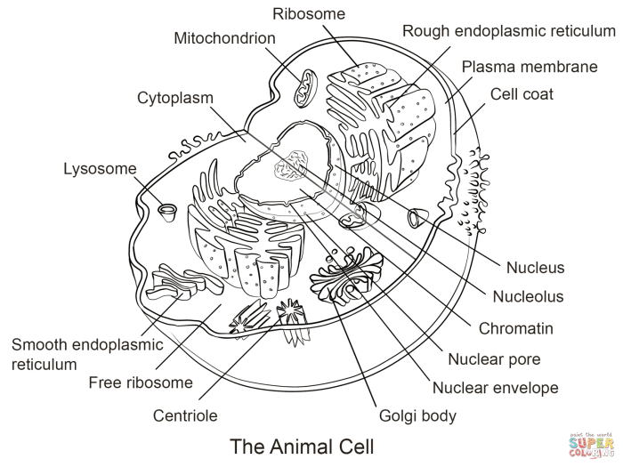
This is the boss level. We’re going all out here. Every organelle is represented, with detailed shapes and colors to distinguish them. The nucleus (a rich, deep purple with a visible nucleolus in a lighter shade of purple), is clearly the center of attention. The mitochondria (bright orange, with internal cristae subtly indicated by shading), endoplasmic reticulum (a more intricate green network, with clear differentiation between rough and smooth ER), Golgi apparatus (pale yellow, with clearly defined cisternae), lysosomes (small, dark red circles), ribosomes (tiny, dark blue dots scattered throughout the cytoplasm), and the centrosome (a pair of centrioles, represented as small, cylindrical structures in a light pink) are all meticulously shown.
We can even add vacuoles (small, light blue circles) and peroxisomes (small, light green circles). Using a combination of color and shading creates a 3D effect that further enhances the visual appeal and understanding of the cell’s intricate structure. For instance, using lighter shades of colors to represent the interior of the organelles creates depth. The color scheme should be consistent but varied enough to distinguish different components without visual overload.
Comparison of Coloring Styles and Complexity
The effectiveness of different coloring styles boils down to clarity and ease of understanding. Simple, consistent colors are best for basic illustrations, while more complex illustrations can benefit from a wider range of colors and shading techniques to highlight the different organelles and their functions.
- Simple Illustration: Minimal organelles, basic shapes, uniform coloring. Easy to understand, but lacks detail.
- Intermediate Illustration: More organelles, slightly more complex shapes, color-coding introduced. Balances detail and clarity.
- Complex Illustration: All major organelles, intricate shapes, varied colors and shading. Highly detailed, but may be overwhelming for beginners.
Popular Questions: Labeled Animal Cell Coloring
What are some common mistakes to avoid when coloring an animal cell?
Common mistakes include using colors that are too similar, neglecting to label organelles clearly, and not accurately representing the relative sizes and shapes of organelles.
How can I make my labeled animal cell diagram more visually appealing?
Use a variety of colors, add shading for depth, and consider using different textures or patterns to represent different organelles. Neatness and clear labeling are also key.
Are there online resources that can help with labeled animal cell coloring?
Yes! Many websites and educational platforms offer printable diagrams, interactive activities, and tutorials on animal cell structure and coloring techniques.
What are the best types of coloring tools to use for this activity?
Colored pencils, markers, or crayons work well, depending on your preference and desired level of detail. Fine-tipped markers are good for precise labeling.

